Dr Evelyn Tay is a MOH-accredited Consultant Dermatologist with over 15 years of experience, specialising in Mohs surgery, skin cancer treatment, aesthetics and laser therapy.
Small bumps around the eyes can be surprisingly difficult to identify. Milia, syringomas, sebaceous hyperplasia and several other benign skin growths can look similar from a distance, yet they arise from different structures in the skin and respond to different treatments.
Syringomas are especially common around the lower eyelids. They are harmless, usually painless and do not turn into skin cancer. Treatment is generally cosmetic, but removing them is less straightforward than treating a superficial blocked pore. Syringomas arise in the dermis, so treatment must reach deep enough to reduce the lesion while protecting surrounding skin from unnecessary scarring or pigment change.
A syringoma is a benign growth that develops from cells associated with an eccrine sweat duct. Eccrine glands occur throughout the skin and produce sweat, while syringomas arise from the ductal portion of this system. They usually form small, firm papules rather than large lumps.
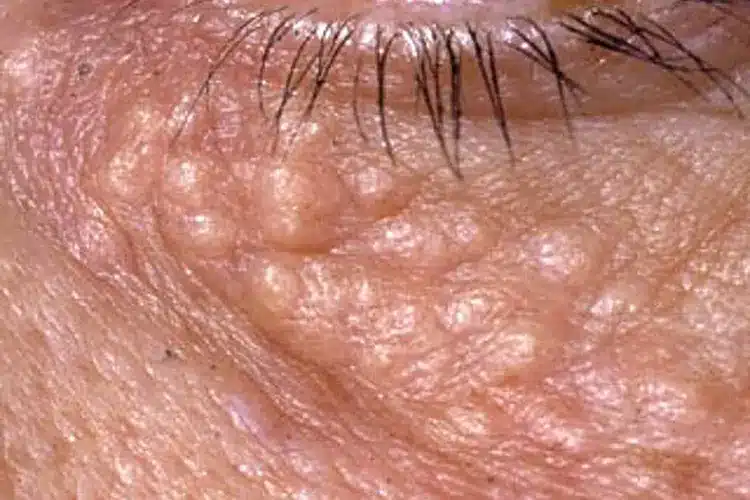
Most syringomas measure around 1 to 3 mm and occur in clusters. The lower eyelids and upper cheeks are the classic locations, often with a similar pattern on both sides of the face. Syringomas can also appear on the forehead, neck, chest, underarms and genital skin, although these locations are less common.
The lesions remain benign. Their main impact is cosmetic, particularly when numerous bumps change the texture of the delicate skin around the eyes. Some people notice only a few lesions, while others gradually develop larger clusters over several years.
What Causes Syringomas
Syringomas develop when cells within the eccrine sweat duct proliferate and form a small benign tumour within the skin. The exact reason this happens in an individual patient is often unclear. They should not be thought of as blocked pores, trapped sweat or acne, and cleansing more aggressively does not prevent them.
Genetics can contribute. Familial syringomas occur in some families and may appear earlier in life. Hormonal influence has also been proposed because syringomas commonly emerge around adolescence or early adulthood and occur more frequently in women. Certain forms have recognised associations with conditions such as Down syndrome, although most people with ordinary eyelid syringomas have no associated medical disorder.
Eruptive syringomas form a less common pattern in which numerous papules appear over a wider area, such as the neck, chest, abdomen or limbs. This variant is reported more often in Asian and darker-pigmented skin. Its broader distribution can make diagnosis less obvious than the familiar clusters around the eyelids.
What Syringomas Look and Feel Like
Typical syringomas are firm, rounded bumps with a smooth surface. They may be skin-coloured, cream, pale yellow, slightly translucent or more pigmented than the surrounding skin. Around the eyes, they often form several small papules below the lower eyelid rather than one isolated growth.
Most cause no physical symptoms. They usually do not hurt, become inflamed, or discharge material when pressed. Occasional itching can occur, particularly with sweating or in certain anatomical locations. A sudden painful or inflamed bump around the eye is therefore less typical of an ordinary syringoma and may represent another condition.
Appearance can vary with skin tone. In more deeply pigmented skin, syringomas may appear yellow-brown, lighter than the surrounding skin or distinctly hyperpigmented. Their small size and uniform clustering can provide a clue, but visual appearance alone is not always enough for a confident diagnosis.
Syringoma Milia and Other Small Facial Bumps
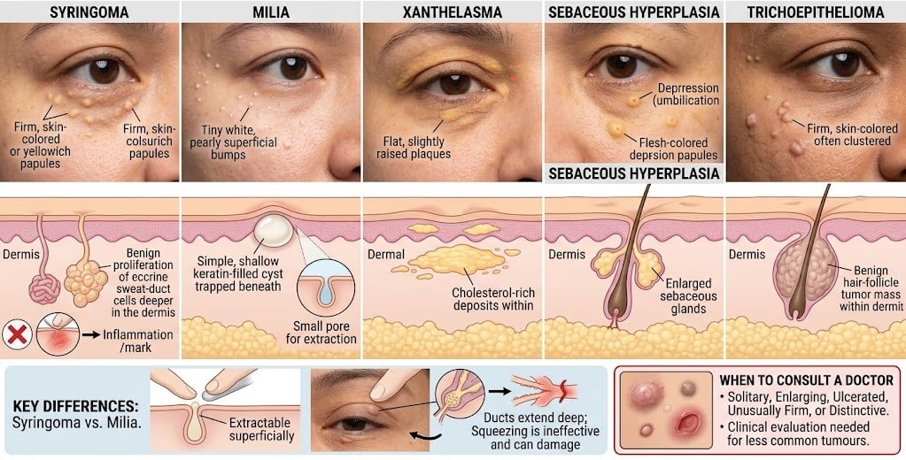
One reason people try ineffective home treatments is that several conditions around the eyes look alike. The structure underneath the bump determines which treatment can work.
Condition
Typical appearance
What forms the lesion
Syringoma
Multiple firm, skin-coloured or yellowish papules, commonly under the eyes
Benign proliferation of eccrine sweat-duct cells
Milia
Tiny white or pearly superficial bumps
Keratin trapped beneath the skin surface
Xanthelasma
Flat or slightly raised yellow plaques around the eyelids
Cholesterol-rich deposits within the skin
Sebaceous hyperplasia
Yellowish or flesh-coloured papules, often with a central depression
Enlarged sebaceous glands
Trichoepithelioma
Firm skin-coloured papules, sometimes multiple
Benign hair-follicle tumour
Milia are typically situated closer to the skin surface and may be extractable through a small pore. Syringomas are different because their ductal structures extend deeper into the dermis. Trying to squeeze one usually produces nothing and can leave inflammation or a mark.
Some less common tumours can also resemble syringomas clinically. A lesion that is solitary, enlarging, ulcerated, unusually firm or different from the surrounding papules deserves closer examination rather than automatic cosmetic treatment.
How a Dermatologist Diagnoses Syringoma
Dermatologists can often recognise typical eyelid syringomas during a clinical examination. The dermatologist considers their size, colour, distribution, depth and symmetry, along with how long they have been present and how they have changed.
A biopsy may be appropriate when the appearance is atypical, or another tumour needs to be excluded. Histology shows characteristic ductal structures within the dermis. The pathologist may describe small ducts lined by epithelial cells, sometimes with comma-shaped extensions that create a distinctive microscopic appearance.
Diagnosis should come before removal when there is uncertainty. Treating every small facial bump as syringoma can lead to unnecessary procedures, while assuming an unusual lesion is benign may delay the diagnosis of another condition.
How to Get Rid of Syringoma
There is no medical requirement to remove an ordinary syringoma. Patients usually choose treatment because the bumps affect the smoothness or appearance of the eyelid or face. The main challenge is their depth. Removing too little tissue can leave enough syringoma behind for it to remain visible or recur, while treating too aggressively increases the risk of scarring or pigment change.
No single technique is ideal for every patient. Lesion number, depth, location, skin tone, previous treatment, and scarring tendency all influence the choice. Multiple conservative sessions can sometimes provide a better cosmetic balance than attempting complete removal in one aggressive treatment.
Common procedural approaches include the following.
Carbon Dioxide Laser
CO2 laser can precisely ablate small areas of tissue and is an established option for syringomas. The dermatologist controls treatment depth lesion by lesion when treating clusters around the eyes.
Healing commonly involves temporary redness, crusting and swelling. Post-inflammatory hyperpigmentation can occur, particularly in more pigmented skin, and excessive treatment depth can cause scarring. Treatment therefore aims for controlled reduction rather than indiscriminate destruction of the entire area.
Electrosurgery
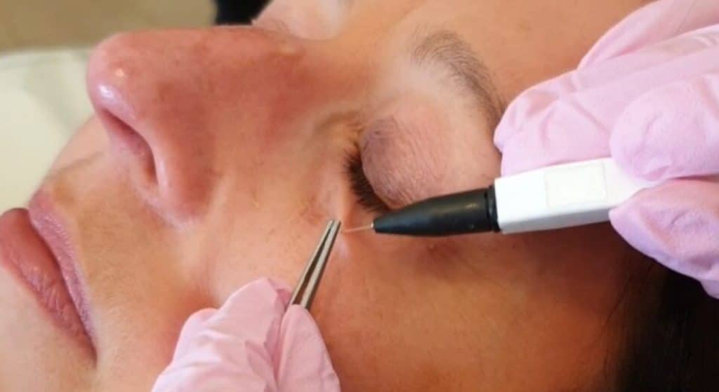
Electrosurgery uses electrical energy to create controlled heat within the lesion. A fine treatment tip allows individual papules to be treated with limited disruption to neighbouring skin.
As with laser treatment, technique and depth influence both clearance and cosmetic outcome. Small crusts can form during healing, and multiple treatments may be appropriate for numerous or deeper syringomas.
Surgical Removal
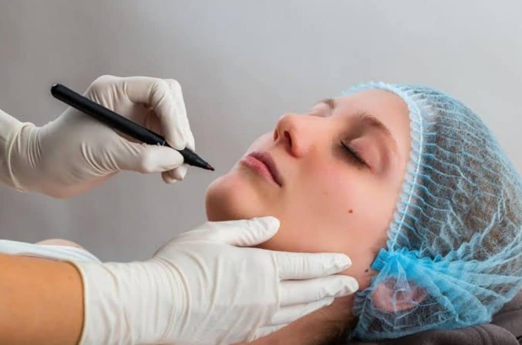
Individual syringomas can sometimes be removed surgically, although this approach is less practical for dozens of tiny eyelid papules. Any incision creates a scar, so surgery tends to have a narrower role than targeted laser, electrosurgery or radiofrequency treatment.
Excision may be more relevant when the diagnosis is uncertain, and tissue is needed for histopathological examination. In that situation, diagnosis takes priority over purely cosmetic removal.
How to Remove SyringomaAround the Eyes
Periorbital syringomas need particular care because eyelid skin is thin and visually unforgiving. Even a small depression, pigment change or scar can become noticeable in this area. The treatment plan therefore needs to account for lesion depth as well as the amount of healthy tissue between neighbouring papules.
A dermatologist may treat individual lesions conservatively and allow them to heal before deciding how much further treatment is needed. Closely packed syringomas do not always need to disappear completely for the skin texture to look substantially smoother. Preserving the eyelid's natural contour and colour can matter more than pursuing complete clearance at any cost.
Eye protection is also required for procedures that deliver laser or energy close to the eye. This is another reason home devices and non-medical attempts at eyelid removal are inappropriate.
Syringoma Treatment in More Pigmented Skin
Syringomas are common in Asian skin, and treatment planning should account for post-inflammatory pigment changes. Heat, ablation, and inflammation can leave temporary darkening after a procedure. Less commonly, excessive tissue injury can result in lighter areas or scarring.
Treatment settings therefore need to balance lesion depth against the skin's response to injury. A staged approach may be preferable when there are many lesions or a history of post-inflammatory hyperpigmentation. Sun protection during healing also reduces additional pigment stimulation.
Previous reactions to lasers, peels, wounds, or acne can give the dermatologist information about how your skin tends to heal. Mention any history of keloids, prolonged pigmentation or unusual scarring before treatment.
Why Syringomas Can Return After Removal
Syringomas extend into the dermis. If treatment only removes the visible surface, deeper ductal structures may remain, and the papule can become noticeable again. New syringomas can also develop nearby over time.
Recurrence does not necessarily mean the original procedure was performed incorrectly. Complete destruction of every deep component would require more tissue injury and may produce a worse cosmetic result than accepting a degree of recurrence. Treatment therefore involves a deliberate balance between clearance and preservation of normal skin.
Patients with numerous lesions may need more than one session, especially around the eyelids. Follow-up allows the dermatologist to assess which papules have flattened sufficiently and which can safely receive additional treatment.
Why Creams and Home Removal Usually Do Not Work
Established syringomas are dermal growths, not clogged pores. Acne creams, salicylic acid products, exfoliating scrubs and pore strips do not physically remove the underlying ductal proliferation. Topical retinoids may sometimes soften the appearance of very small lesions, but they rarely remove established syringomas completely.
Cutting, squeezing or burning the bumps at home creates far greater risk than benefit. The eyelids are particularly prone to visible scars and post-inflammatory pigment changes. Strong acids purchased online can also cause chemical burns without reaching the lesion in a controlled way.
If a bump has never been professionally diagnosed, home treatment introduces another problem, since milia, syringomas, sebaceous growths and several other lesions require different approaches, and some uncommon tumours can mimic benign facial papules.
When to Have the Bumps Checked
Clusters of small, stable, symmetrical bumps below both eyes commonly fit the pattern of syringomas. A dermatology consultation can confirm the diagnosis if their appearance bothers you or if you are considering removal.
Assessment becomes more important when a lesion behaves differently from the others. Rapid growth, bleeding, ulceration, pain, significant inflammation or one unusual solitary bump is not the classic syringoma pattern. A dermatologist can decide whether clinical examination is enough or whether a biopsy is appropriate.
A consultation before cosmetic treatment also gives you realistic expectations for clearance, downtime, recurrence, and pigment risk. This is especially relevant when many lesions occupy a large part of the lower eyelid.
How Lumine Dermatology Can Help
Lumine Dermatology & Laser Clinic can assess small bumps around the eyes or elsewhere on the face to confirm if they are syringomas and rule out other conditions that can look similar. The diagnosis guides treatment, particularly around the eyelids where the skin is thin and the risk of visible pigment change or scarring needs careful consideration.
We plan treatment based on the number, depth, and location of the lesions, as well as the patient’s skin tone and previous response to procedures. For patients with multiple periorbital syringomas, treatment may be staged to improve texture while limiting unnecessary injury to the surrounding skin.
Syringomas are harmless, but treating them well requires more than removing the visible bump. The depth of the lesion, the delicate skin around the eyes and the risk of pigmentation or scarring all influence the result.
A careful assessment at Lumine Dermatology can determine the right treatment approach and set realistic expectations for improvement, recovery, and the possibility of recurrence.
Healthy skin, Happy mood.
We've got your back!
Every skin concern matters. Drop an enquiry, and we'll get back to you within one working day.
Address:
1 Scotts Road #04-15/16 Shaw Centre Singapore 228208
Operation hours
Mon-Fri - 9am to 6pm | Sat - 9am to 1pm | Closed on Sundays & Public Holidays