Squamous Cell Carcinoma: Skin Cancer Warning Signs
Article medically reviewed by Dr. Evelyn Tay
Dr Evelyn Tay is a MOH-accredited Consultant Dermatologist with over 15 years of experience, specialising in Mohs surgery, skin cancer treatment, aesthetics and laser therapy.
A persistent rough patch, a firm growth, or a sore that repeatedly crusts over can indicate early squamous cell carcinoma. These lesions may resemble common skin problems, particularly when they are small or cause little discomfort. Changes in skin texture and how a lesion develops over time often raise the first concern.
Squamous cell carcinoma (SCC) is a common form of skin cancer that usually develops on areas exposed to sunlight over many years. Most cases respond well to treatment when diagnosed early, although some tumours can grow into deeper tissue or spread to nearby lymph nodes.
Recognising suspicious changes allows patients to seek assessment before a lesion becomes more extensive. The location, growth pattern and microscopic characteristics of the tumour all influence the treatment a dermatologist may recommend.
The outer layer of the skin contains keratinocytes, cells that produce keratin and contribute to the skin's protective structure. Squamous cell carcinoma develops when abnormal keratinocytes multiply without normal growth control, forming a malignant lesion. As the tumour becomes invasive, cancer cells extend beyond the epidermis into deeper skin tissue.
Most cutaneous SCCs develop gradually, although some grow considerably faster. A lesion may remain relatively small on the surface while extending into the surrounding skin. This behaviour explains why treatment decisions cannot rely on visible size alone.
Among the main skin cancer types, SCC differs from basal cell carcinoma and melanoma in its cellular origin and typical behaviour. Basal cell carcinoma rarely spreads beyond its original site, while melanoma arises from pigment-producing cells and carries a different risk profile. Squamous cell carcinoma can invade surrounding structures and occasionally spread elsewhere, particularly when certain high-risk features are present.
RecognisingSquamous Cell Carcinoma Symptoms
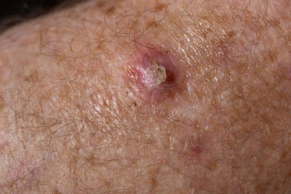
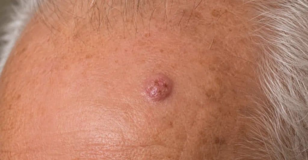
The appearance of SCC varies considerably. Some lesions remain relatively flat and develop a thickened, scaly surface. Others form raised nodules with a firm base or a central area of crusting. A persistent growth that resembles a wart may also require examination, especially when it enlarges or becomes tender.
Texture changes can be more noticeable than colour changes. Skin that develops a hard, rough or thickened area without an obvious cause may need assessment, particularly if ordinary moisturising or other basic care makes little difference.
The following appearances deserve closer attention:
Skin change
Features that may raise concern
Persistent scaly patch
A rough area that gradually thickens, becomes more defined or develops a firm underlying texture
Raised nodule
A solid, sometimes dome-shaped growth that continues enlarging
Recurrent crusting
A lesion that repeatedly forms a scab, sheds it and develops another
Non-healing ulcer
An open area that persists, bleeds or develops raised edges
Wart-like growth
A rough, projecting lesion that becomes larger, harder or more irregular
Tender or painful spot
A persistent lesion that develops increasing discomfort or altered sensation
Early squamous cell carcinoma symptoms may be limited to one small area. Bleeding and pain are possible, but neither needs to be present for a lesion to be malignant. The combination of persistence, progressive growth and structural change usually provides a stronger reason for assessment than any isolated symptom.
Changes That Deserve Prompt Medical Attention
A growth that becomes noticeably thicker or larger over several weeks warrants examination, particularly if it has developed on previously sun-damaged skin. Repeated bleeding without a clear injury, spontaneous ulceration and persistent tenderness also require investigation.
Pain deserves particular consideration when it develops within a known or suspected SCC. Most early lesions do not cause substantial discomfort, but persistent pain, numbness, or tingling around a tumour can occasionally indicate nearby nerve involvement. These symptoms require prompt specialist assessment rather than repeated treatment with ordinary skincare products.
An unexplained lump in a nearby lymph node region is another reason to seek medical attention. For example, certain cancers on the scalp, face or ear may spread to lymph nodes around the head and neck. This is uncommon in many early cases, but the possibility influences examination and treatment planning for higher-risk tumours.
How Sun-Damaged Skin Can Develop Into SCC
Long-term ultraviolet exposure can damage DNA in keratinocytes. Over time, affected cells may develop abnormal growth patterns, leading to precancerous lesions or skin cancer. The amount of accumulated damage often reflects many years of exposure rather than one recent episode of sunburn.
Actinic keratoses are an example of this process. They commonly develop as rough, scaly areas on the face, ears, scalp, forearms and backs of the hands. Some feel like a small patch of sandpaper before they become particularly visible. Although most actinic keratoses do not develop into invasive cancer, a proportion can progress to SCC.
Dermatologists also distinguish actinic keratosis from squamous cell carcinoma in situ, sometimes called Bowen's disease. In SCC in situ, malignant cells remain confined to the epidermis. In invasive SCC, malignant cells extend beyond that outer layer into deeper tissue.
These distinctions affect management. A suspicious thickened lesion may require biopsy to establish if abnormal cells remain superficial or have become invasive. Repeatedly freezing or applying topical treatments to an undiagnosed lesion can delay recognition of a deeper tumour.
WhereSquamous Cell CarcinomaCommonly Develops
Areas exposed to sunlight are frequent locations for cutaneous SCC. The face, ears, lips, neck, scalp, forearms and backs of the hands receive considerable cumulative UV exposure, especially in people who spend substantial time outdoors.
Certain locations also require more careful treatment consideration. For example, tumours on the ear or lip may have a higher risk of recurrence or spread than small, uncomplicated lesions elsewhere on the body. Skin cancers near the eyelids, nose and other anatomically delicate areas require treatment that preserves normal function and surrounding tissue.
SCC can also occur on skin that receives limited sunlight. Long-standing wounds, burn scars and areas of chronic inflammation occasionally develop malignant changes. Persistent ulceration or new growth within an old scar should therefore be examined.
Squamous Cell CarcinomaAcross Different Skin Tones
People with fair skin have a higher overall risk of UV-related SCC because they possess less natural protection against ultraviolet damage. However, people with Asian and darker skin tones can also develop this form of cancer.
Appearance can differ across complexions. A lesion may look pink or red on lighter skin, while some growths appear brown, grey, darker than the surrounding skin or close to the patient's natural skin colour. Texture, firmness, surface crusting and growth pattern remain relevant regardless of pigmentation.
In more deeply pigmented skin, SCC may also develop with chronic wounds, scars, or inflammation rather than appearing exclusively in heavily sun-exposed areas. A skin change should therefore be assessed according to its behaviour and clinical characteristics, without assuming that darker skin provides complete protection against skin cancer.
Who Has a Higher Risk of Developing SCC?
Cumulative ultraviolet exposure is a major contributor to cutaneous SCC. People who spend years working or exercising outdoors may accumulate considerable skin damage, especially when sun protection has been inconsistent. Previous actinic keratoses or skin cancers can indicate that the skin has already experienced substantial UV-related change.
Immune suppression creates a different risk profile. Organ transplant recipients and people taking certain long-term immunosuppressive medicines can develop SCC more frequently, and some tumours may behave more aggressively. A history of radiotherapy, chronic wounds or significant burn scarring can also influence susceptibility.
These factors affect clinical decisions after a suspicious lesion appears. Someone with a previous SCC or a suppressed immune system may need closer skin surveillance and more urgent investigation of a new growth. The dermatologist considers individual medical history alongside the lesion's appearance rather than treating every suspicious spot as having the same level of risk.
How a Dermatologist DiagnosesSquamous Cell Carcinoma
An assessment begins with the lesion's history and physical characteristics. The dermatologist examines its surface, borders, firmness and relationship to the surrounding skin, while considering how long it has been present and how quickly it has changed.
Dermoscopy can reveal structural and vascular features that are difficult to assess with ordinary inspection. Certain patterns may increase suspicion of SCC, although dermoscopy cannot establish a definitive diagnosis or reliably determine the full depth of a tumour.
When the findings raise concern, a skin biopsy provides tissue for microscopic examination. Depending on the lesion's size, location and appearance, the dermatologist may take a sample using a shave or punch technique or remove the entire growth.
The pathologist examines the tissue for malignant keratinocytes and evidence of invasion. The report can also describe features such as tumour differentiation, depth and possible involvement of nerves or other surrounding structures. These findings influence treatment choice and the need for further investigation.
For selected higher-risk tumours, the examination may extend to nearby lymph nodes. Imaging or further specialist assessment may be appropriate if the clinical findings suggest deeper involvement or spread. Routine scans are generally unnecessary for uncomplicated early SCC.
Why the Biopsy Result Influences Treatment
Two lesions with a similar surface appearance can require different treatment plans. A small, superficial tumour with clearly defined margins may be straightforward to remove. A tumour with poorly defined borders, greater depth or aggressive microscopic features may require more extensive assessment and surgery.
The pathology report provides information that cannot be established from photographs or the surface appearance alone. Poor differentiation, perineural invasion and other concerning findings can indicate a greater risk of recurrence or spread. Location, tumour size and the patient's immune status add further context.
This risk assessment influences how much surrounding tissue needs examination, the type of surgery recommended and the follow-up schedule after treatment. Patients with higher-risk disease may also need care from additional specialists to assess regional lymph nodes or plan further treatment.
Squamous Cell Carcinoma TreatmentOptions
For most invasive cutaneous SCCs, surgical removal remains the principal treatment. The aim is to remove the cancer completely and confirm that the surrounding tissue margins are free of malignant cells. The method depends on tumour characteristics, not a single procedure suitable for every case.
Surgical Excision
Standard excision involves removing the tumour together with an appropriate margin of surrounding skin. The dermatologist or surgeon then closes the wound, where appropriate, and sends the tissue for histopathological examination.
Microscopic analysis determines if the margins are clear. If cancer cells remain at the edge of the specimen, further treatment may be necessary. The size and location of the defect determine the closure technique and how the area is expected to heal.
Mohs Micrographic Surgery
Mohs surgery offers a more detailed method of examining surgical margins. The surgeon removes the tumour in stages and examines the tissue microscopically, continuing until the assessed margins no longer contain cancer cells.
This approach may be particularly appropriate for selected SCCs on the face, ears, lips or other areas where conserving healthy tissue is a priority. Mohs surgery may also benefit recurrent tumours, poorly defined lesions, and certain higher-risk cancers.
Because Mohs surgery combines precise tissue removal with detailed margin assessment, it can reduce the amount of healthy skin sacrificed while supporting complete tumour clearance. The technique still requires careful wound reconstruction, and a scar remains after healing.
When Other Treatments May Be Considered
Not every diagnosis requires the same surgical approach. Selected cases of SCC in situ may be treated with topical medication or other local procedures, depending on their size, location and clinical characteristics. These methods have a narrower role in invasive disease because they may not provide the same degree of tumour clearance or margin assessment as surgery.
Curettage with electrodesiccation may be appropriate for certain carefully selected low-risk superficial tumours. Radiotherapy can provide another option when surgery is unsuitable or, in selected cases, as part of additional treatment after an operation.
For advanced SCC that surgery or radiotherapy cannot adequately treat, specialist oncology care may include systemic immunotherapy. These treatments target cancer-related immune pathways and require assessment of disease extent, general health, and potential side effects.
An individual squamous cell carcinoma treatment plan therefore depends on the confirmed diagnosis, tumour risk and available procedures. Early-stage disease often requires a much more limited intervention than a tumour that has invaded deeper structures or spread.
Recovery and Follow-Up After Treatment
Recovery depends on the treatment site, the amount of tissue removed and the closure method. A small excision on the arm may heal differently from a larger surgical wound near the nose or eyelid. Patients receive instructions on wound cleaning, dressing care, physical activity and when to return for review or suture removal.
The final pathology result remains part of postoperative care. It confirms the diagnosis and provides information about tumour clearance and any additional risk features. If the findings suggest incomplete removal or unexpectedly aggressive disease, the treatment plan may need adjustment.
A previous SCC also increases the likelihood of developing another skin cancer. Follow-up examinations therefore look for recurrence near the original treatment area and new suspicious lesions elsewhere. The frequency depends on the tumour's risk profile, the patient's medical history and the findings during subsequent skin checks.
Reducing Skin Cancer Risk in Singapore
Singapore's year-round ultraviolet exposure makes consistent sun protection relevant even for people who spend much of their time indoors. Daily exposure accumulates during commuting, walking, driving, and outdoor activities, particularly on the face, ears, neck, and hands.
Broad-spectrum sunscreen with SPF 30 or higher, protective clothing, hats and shade can reduce avoidable UV exposure. During sustained outdoor activity, apply and reapply sunscreen as needed to maintain protection. People with extensive sun damage or a previous skin cancer may need a more structured prevention and surveillance plan.
Skin checks should also account for growths that do not resemble moles. A persistent scaly lesion, thickening within an old scar or a sore that repeatedly breaks down deserves examination. Photographs can document visible changes over time, although a concerning lesion should not be monitored indefinitely instead of receiving medical assessment.
How Lumine Dermatology Can Help
Lumine Dermatology & Laser Clinic provides specialist assessment and treatment for suspicious skin lesions, including squamous cell carcinoma and other forms of skin cancer. Clinical examination and dermoscopy can help identify lesions that require biopsy, while histopathological findings guide subsequent treatment decisions.
For confirmed skin cancer, management may include surgical excision or Mohs micrographic surgery when appropriate. The approach considers the tumour's location, size, borders and microscopic features, alongside the need to preserve surrounding tissue and function. We can coordinate additional specialist care if the disease requires further investigation or treatment.
Early examination can prevent a persistent skin change from progressing unnoticed. If a growth continues to enlarge, develops a thick crust, bleeds repeatedly or fails to heal, a consultation at Lumine Dermatology can establish the diagnosis and determine the appropriate next step. The aim is accurate cancer removal, careful wound management, and continued surveillance based on the patient's individual risk.
Healthy skin, Happy mood.
We've got your back!
Every skin concern matters. Drop an enquiry, and we'll get back to you within one working day.
Address:
1 Scotts Road #04-15/16 Shaw Centre Singapore 228208
Operation hours
Mon-Fri - 9am to 6pm | Sat - 9am to 1pm | Closed on Sundays & Public Holidays