Dr Evelyn Tay is a MOH-accredited Consultant Dermatologist with over 15 years of experience, specialising in Mohs surgery, skin cancer treatment, aesthetics and laser therapy.
Melanoma does not always begin as a large, dark or obviously abnormal mole. The early signs of melanoma can be much less striking. A mole may slowly develop a different outline, gain a second colour, or become noticeably different from the others on your skin. Melanoma can also appear as a new spot where no mole existed before.
Early recognition depends less on finding a single textbook appearance and more on noticing change. Dermatologists assess the shape, colour, structure and behaviour of a lesion, then compare those features with the rest of the patient’s skin. A suspicious change does not confirm skin cancer, but it gives a clear reason for closer examination.
Many early melanomas contain brown or black pigment, although their colour can vary considerably. A lesion may appear as a flat brown patch, a mole with irregular pigmentation, or a small mark whose outline becomes less even over time. Some contain several shades of brown or black, while others include areas of pink, red, white, grey, or blue.
Early cancerous moles can still be small. A lesion does not need to become large to warrant attention. A change in a 3 or 4 mm mole may be more significant than the size of a stable 8 mm mole that has looked the same for years. Diameter is one feature among several, not a threshold for concern.
Melanoma can also arise on skin that previously looked normal. A new pigmented lesion that persists, enlarges, darkens, or changes in structure should be assessed in the context of the person’s age, other moles, and skin history.
The ABCDESigns of Melanoma
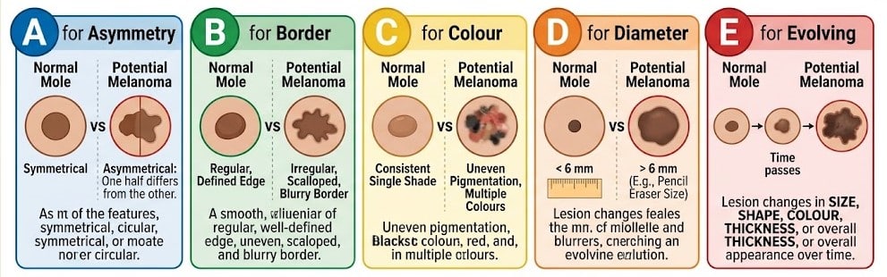
The ABCDE rule provides a structured way to check pigmented lesions for features associated with melanoma. It is particularly helpful when comparing an existing mole with how it looked previously.
Sign
What to look for
A for Asymmetry
One half differs from the other in shape, colour or internal pattern.
B for Border
The edge becomes irregular, blurred, notched or poorly defined.
C for Colour
The lesion contains uneven pigmentation or several colours rather than one consistent shade.
D for Diameter
Melanomas are often larger than 6 mm when diagnosed, although early melanomas can be considerably smaller.
E for Evolving
The lesion changes in size, shape, colour, thickness or overall appearance.
Of these features, evolution often carries particular weight. Benign moles usually remain stable. A lesion that begins changing after a period of stability may need closer assessment even if it does not look striking at a single point in time.
The ABCDE rule also has limits. Some melanomas remain fairly symmetrical, contain one dominant colour, or measure less than 6 mm. Nodular and amelanotic melanomas can look very different from the classic irregular dark mole, so a lesion should not be considered reassuring simply because it does not meet every ABCDE criterion.
The Mole That Looks Different From Your Other Moles
A dermatologist does not assess every mole against one universal picture of what a normal mole should look like. People often develop their own recognisable pattern of naevi. One person may have numerous small, pale-brown flat moles, while another has several darker raised ones. A lesion that breaks that personal pattern can be clinically significant.
Dermatologists often call this the ugly duckling sign. The lesion may be darker, lighter, larger, more irregular, or structurally different from the surrounding moles. It may also be the only lesion that has recently changed. This comparison is especially relevant for people with many moles, where the most concerning spot may be the one that breaks the patient’s usual pattern.
An unusual mole is not automatically melanoma. Atypical naevi and several benign skin growths can differ from neighbouring lesions. The ugly duckling sign helps identify which lesions deserve closer dermoscopic examination rather than attempting to diagnose melanoma from appearance alone.
Nodular Melanoma May Not Follow the Classic ABCDE Pattern
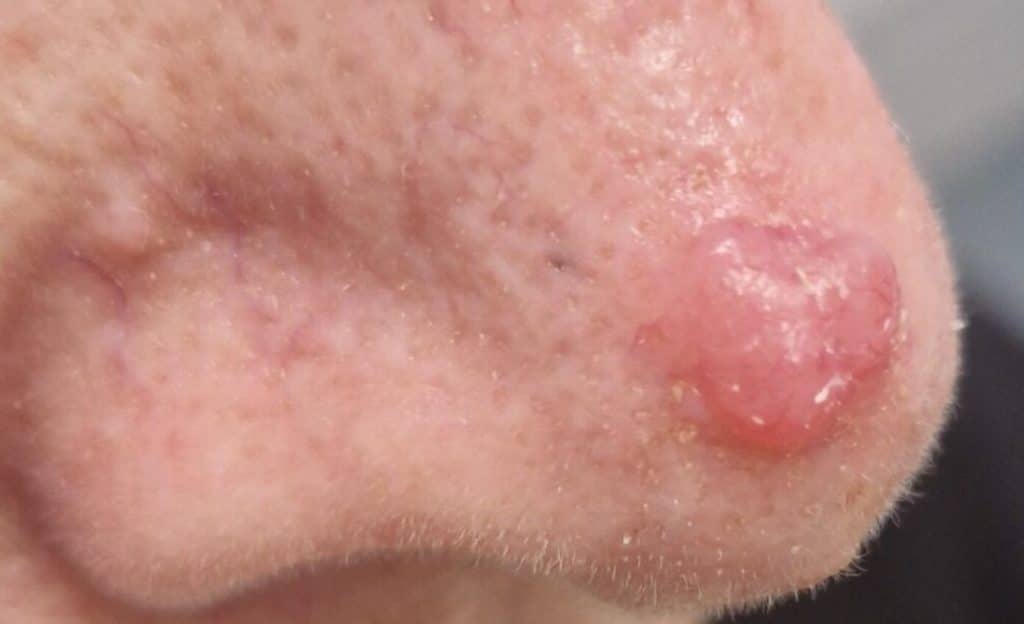
Nodular melanoma deserves separate attention because it can behave differently from the more familiar flat, spreading melanoma. It often develops as a raised growth and may increase in size over a relatively short period. The lesion can be black, brown, red, pink or close to skin colour, and it may have a relatively regular outline.
The EFG rule can help identify this pattern. E stands for elevated. F stands for firm. G stands for growing. A newly developed bump that feels firm and continues to enlarge over weeks should not be dismissed because it lacks several ABCDE features. Bleeding, crusting or ulceration may develop as the lesion progresses, although these are not required for melanoma to be present.
Raised or firm lesions are not automatically melanoma. Cysts, dermatofibromas, benign moles, and inflamed skin growths can look similar. Persistent growth is more concerning, especially when a new nodule keeps enlarging instead of settling.
Melanoma Can Develop Without Dark Pigment
Amelanotic melanoma contains little visible melanin, so it may appear pink, red or skin-coloured rather than brown or black. This removes one of the visual clues people commonly associate with melanoma and can allow the lesion to resemble an inflamed spot, scar, benign growth or persistent pimple.
A pink or skin-coloured lesion should be assessed if it continues to enlarge, becomes progressively raised or firm, repeatedly crusts, or bleeds without clear trauma. Persistence helps distinguish it from many inflammatory spots, which usually change as they resolve.
Because pigment may be minimal or absent, early signs of melanoma in these lesions relate more to progressive growth, firmness, surface change, or repeated bleeding than to colour. Dermoscopy can reveal vascular and structural patterns that are not apparent during ordinary visual inspection.
Melanoma on the Palms, Soles and Nails
Acral melanoma develops on the palms, soles, fingers, toes or beneath the nails. These areas receive less attention during routine self-checks, and a lesion can initially be mistaken for a bruise, callus, friction mark or nail injury. Acral melanoma can occur in any skin tone and represents an especially important melanoma pattern in people with darker skin.
On the sole or palm, an early lesion may appear as an irregular brown or black patch that changes gradually. Its border may become less defined, pigmentation may become uneven, or the area may expand. A persistent dark area without a clear cause deserves assessment rather than repeated treatment as a callus or minor injury.
Nail melanoma can begin as a longitudinal brown or black band running from the base towards the tip of a nail. Many nail pigment bands are benign, particularly in people with darker skin. Features that raise greater concern include a new band affecting one nail, progressive widening, irregular colour or width, and pigment extending onto the skin beside the nail. A history of trauma does not automatically explain a mark that persists or continues to change.
Other Changes That Raise Concern
Progressive growth, spontaneous bleeding, ulceration, repeated crusting, or increasing elevation are more concerning than itch or tenderness alone. A previously stable mole that develops one of these changes should be assessed, particularly if its appearance is also evolving.
Itching or tenderness can occur with melanoma, yet these symptoms are not specific. Benign moles, eczema, seborrhoeic keratoses, and inflamed skin growths can cause similar discomfort. These symptoms become more clinically relevant when they accompany visible change.
A lesion that was assumed to be a pimple, bite, or minor wound also deserves review if it continues enlarging or repeatedly breaks down instead of healing. Persistent behaviour can provide an important clue even when the lesion does not resemble a typical dark mole.
How to Monitor Your Skin More Effectively
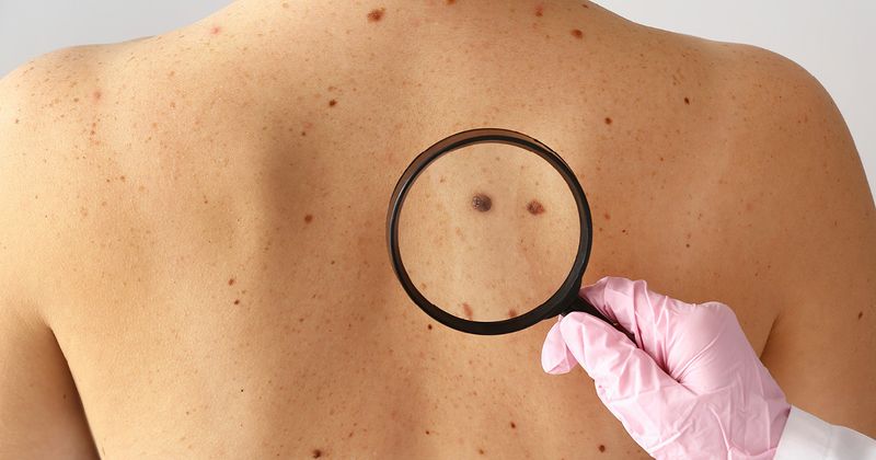
Self-examination works best when you know your skin's baseline appearance. Instead of trying to memorise every mole, look for three categories of change: a new persistent lesion, an existing mole that is evolving, and a lesion that looks different from your usual pattern.
Include areas that are easy to overlook. The scalp, behind the ears, back, buttocks, palms, soles, spaces between the toes and nails all deserve periodic inspection. A mirror or help from someone you trust can make less visible areas easier to check.
Serial photographs can document gradual change when you have numerous moles or a lesion that your dermatologist has advised you to monitor. Use similar lighting, distance, and angle for each image. Placing a ruler beside the lesion can help record size accurately. Photography can support comparison over time, but suspicious changes still require clinical and dermoscopic assessment.
Who May Need Closer Mole Surveillance
Closer surveillance may be appropriate for people with a previous melanoma, a strong family history of melanoma, multiple atypical naevi, a very high mole count, significant UV damage, or immune suppression. These factors do not determine if a particular lesion is melanoma, but they can lower the threshold for investigating new or changing spots.
A patient with a previous melanoma, for example, may need a subtle new lesion assessed sooner than someone with no additional risk factors. People with many atypical moles may benefit from baseline clinical photography or mole mapping, as comparison over time can reveal changes that are difficult to detect during an isolated examination.
The aim of risk assessment is to set an appropriate surveillance plan. It does not replace examination of the individual lesion, and melanoma can still occur in people without obvious risk factors.
What Happens During a Dermatology Mole Assessment
A dermatologist first examines the lesion in the context of the rest of your skin. Its history is important. When did you first notice it? Has it become larger, darker, raised or structurally different? Has it started bleeding or producing symptoms? Previous photographs can be particularly informative if they show genuine evolution.
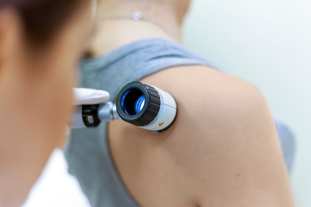
Dermoscopy allows the dermatologist to examine pigment networks, vascular structures, and other patterns beneath the surface of the lesion. The examination is non-invasive and can reveal features that are not visible to the naked eye. These findings help determine if a lesion can be monitored or if tissue examination is needed.
When histopathology cannot exclude melanoma, it provides the definitive diagnosis. Where appropriate, the suspicious lesion is removed and examined microscopically. If invasive melanoma is confirmed, the pathology report includes features such as Breslow thickness, which measures how deeply melanoma cells have entered the skin. Tumour thickness and other histological findings help guide the next stage of treatment.
Why Early Assessment Changes the Treatment Pathway
Melanoma begins in the skin before it can invade deeper and spread. Melanoma in situ remains confined to the epidermis. Once melanoma becomes invasive, depth becomes an important part of staging and treatment planning.
This is why early recognition has direct clinical value. A subtle melanoma can be easier to dismiss than an advanced tumour, yet diagnosis before deeper invasion can lead to a less complex treatment pathway. Waiting for significant bleeding, pain, or obvious enlargement can allow the lesion to progress before assessment.
Early assessment can clarify both outcomes. Suspicious lesions can move to biopsy promptly, while benign lesions can be documented or monitored without unnecessary procedures.
When to Arrange a Mole Check
Arrange a dermatology assessment if an existing mole changes in shape, colour, size, or thickness, or if a new lesion persists and continues to evolve. A spot that looks clearly different from your other moles also deserves attention, even if it does not meet every part of the ABCDE rule.
A growing firm nodule, unexplained pigmentation on the palm or sole, a changing dark band beneath a nail, or a lesion that repeatedly bleeds, crusts, or fails to heal should also be checked. These changes can have benign causes, but appearance alone cannot reliably distinguish them from melanoma.
You do not need to wait for several warning signs to appear at once. A single persistent change can justify assessment, particularly if the lesion is new or behaves differently from the rest of your skin. Recognising the early signs of melanoma means watching for changes, not waiting for a mole to become obviously abnormal.
How Lumine Dermatology Can Help
Lumine Dermatology & Laser Clinic provides specialist assessment for changing moles, unusual pigmented lesions, and suspected skin cancers. Our dermatologists are experts in skin cancer, advanced dermatologic surgery, and Mohs micrographic surgery.
Assessment may include clinical examination, dermoscopy, and comparison with previous photographs where available. If a lesion has concerning features, your dermatologist can arrange biopsy or complete excision for histopathological diagnosis. This helps determine which lesions can be monitored and which require tissue diagnosis or definitive removal.
If you have noticed a new or changing lesion, an early dermatology assessment can provide clarity before the change becomes more advanced. You can arrange a consultation at Lumine Dermatology for mole assessment, dermoscopy, and further investigation where needed.
Healthy skin, Happy mood.
We've got your back!
Every skin concern matters. Drop an enquiry, and we'll get back to you within one working day.
Address:
1 Scotts Road #04-15/16 Shaw Centre Singapore 228208
Operation hours
Mon-Fri - 9am to 6pm | Sat - 9am to 1pm | Closed on Sundays & Public Holidays